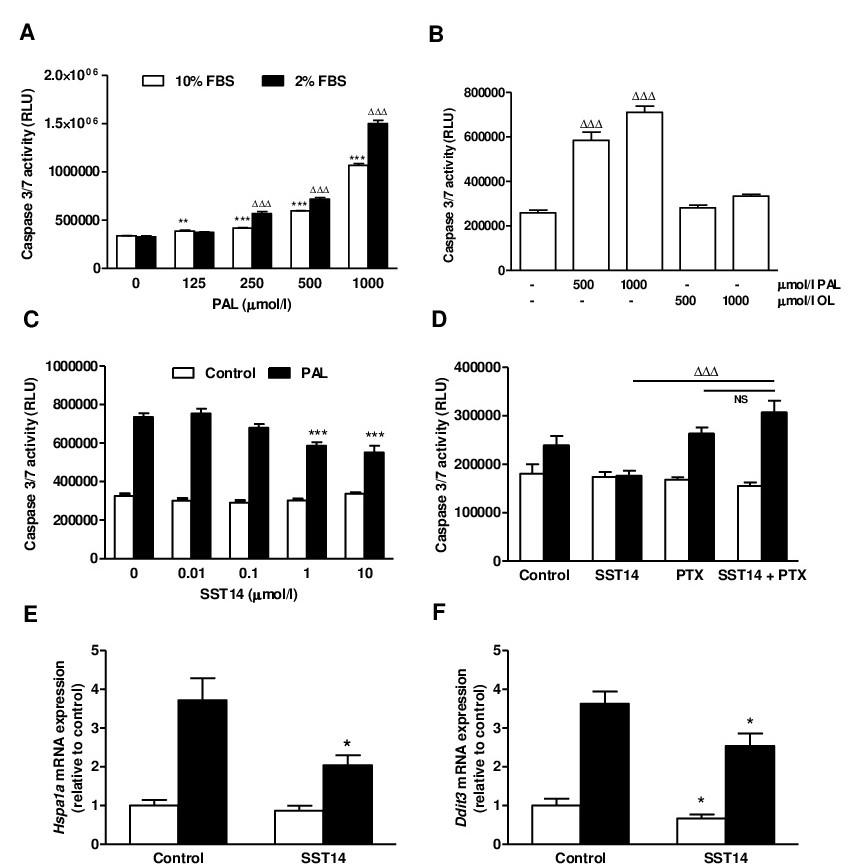
Fig. 1. MIN6 beta cell survival following exposure to palmitate with or without SST14. To optimise the palmitate concentration used for assessment of apoptosis MIN6 beta cells were initially incubated for 20h with increasing concentrations of palmitate (PAL) in DMEM containing 2 or 10% FBS as indicated (panel A). In a separate set of experiments MIN6 cells were exposed to 0.5 or 1 mmol/l palmitate or oleate (OL), respectively, with 2% FBS (panel B). Based on the results, a concentration of 0.5 mmol/l palmitate was subsequently used to induce beta cell apoptosis. MIN6 beta cells were pre-treated for 48h with SST14 followed by 20h incubation with or without palmitate (PAL, black bars) and apoptosis following treatment was assessed by measurement of 3/7 caspase activity (panel C). The observed reduction in apoptosis following SST14 (1 μmol/l) pre-treatment was reversed by co-incubation with the Gi/o inhibitor, pertussis toxin (PTX, 100 ng/ml, panel D). mRNA levels of Hspa1a (panel E) and Ddit3 (panel F) were assessed by qPCR as an indication of cellular and ER stress, respectively, and expression of both were significantly reduced by SST14. Panel A and B: **P<0.01, ***P<0.001 vs 10% FBS control or ∆∆∆P<0.001 vs 2% FBS control, n=8. Panel C-F: *P<0.05, ***P<0.001 vs control without SST14, ∆∆∆P<0.001 SST14 vs SST14 + PTX (one way ANOVA and Bonferroni's multiple comparisons test (A-D) or Student's t test (E and F)). Panel C and D representative of 3-7 experiments. Panel E and F: mean ± SEM of 4-6 separate experiments.